

Meniere's Disease
( Endolymphatic Hydrops ) Meniere's Disease is a disorder characterized by dizziness and/or vertigo, hearing loss, ringing in the ear, and/or a feeling of fullness in the ear.Overview of Meniere's Disease
In 1861, Prosper Meniere described Meniere's disease as a disorder characterized by a constellation of symptoms that include fluctuating hearing loss, a sensation of fullness in the ear, roaring or ringing in the affected ear, and episodic vertigo. There is usually a low- tone hearing loss that fluctuates unpredictably. A sensation of fullness in the ear or a sensation that the ear is clogged is also not uncommon. The roaring tinnitus is usually of a low-tone quality typically described as "ocean waves crashing along the beach.”
Vertigo is a sensation that objects are spinning around you or that you are physically being spun around them. These spells come about at unpredictable times with no warning and last from several hours to several days. It is not uncommon for these spells to be associated with nausea and vomiting. The frequency of these attacks can vary from once a year to three times a day.
All of these symptoms do not necessarily need to be present. Atypical presentations are common.
Atypical Meniere’s Disease
When only some of the symptoms of Meniere’s disease are present, the condition is referred to as atypical Meniere’s disease. For example, the episodes of vertigo may occur without any hearing abnormality or vice versa.
Pertinent Anatomy
Before we embark on further discussion, it is prudent to review and understand pertinent anatomy of the hearing and balance system.
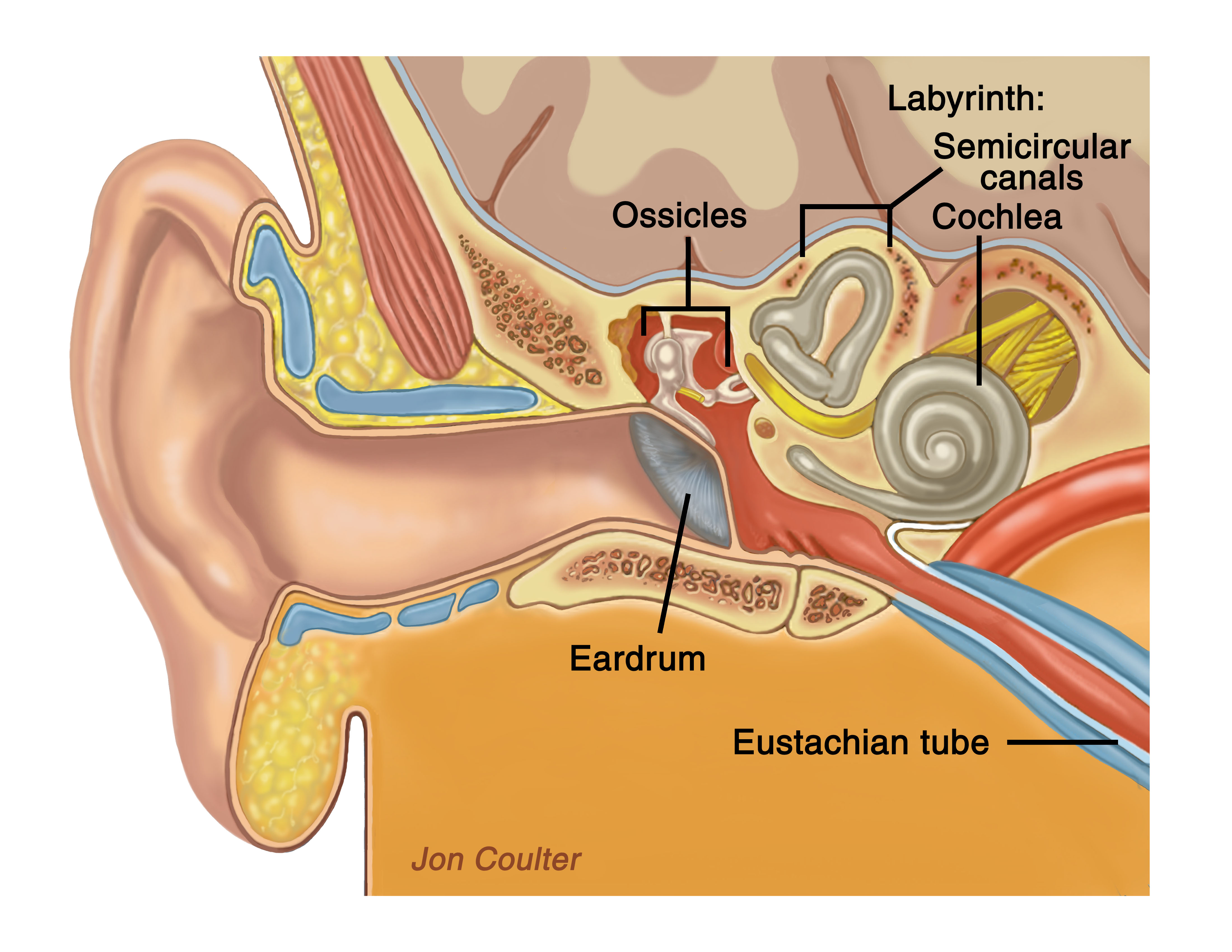
The temporal bone anatomy is shown, including the ear canal, ear drum, middle ear, and inner ear.
Sound entering the ear canal causes vibrations of the tympanic membrane (eardrum). The eardrum is attached to one of three ossicles (bones) found in the middle ear, called the malleus. The malleus bone transmits the mechanical vibrations to another ossicle, the incus, which in turn transmits the vibrations to the smallest of the three ossicles, the stapes. As the stapes vibrates, the fluid in the inner ear is set into motion. The cilia (hairs) on the ends of hair cells within the inner ear are bent, and an electrical signal is generated. This travels along the cochlear (hearing) nerve and then back to the brain. The inner ear is composed of the cochlea, which is responsible for hearing, and the semicircular canals, which convey balance information back to the brain concerning angular acceleration of the head. Two other organs, the saccule and utricle, are found in the inner ear and convey information on linear acceleration of the head.
The internal auditory canal is the bony canal that encases the hearing and balance nerves on their way from the inner ear to the brain. Joining theses nerves in the canal is the facial nerve, which is responsible for allowing motion on the side of the face that the nerve is on. This is the nerve that is responsible for our ability to raise our eyebrows, close our eyes, flare our nostrils, and raise the corner of the mouth on that side. It is also responsible for transmitting information regarding taste from the front 2/3 of the tongue and for producing tears from the lacrimal glands in the eyes.
The inner ear (cochlea and semicircular canals) is composed of two types of fluid-- endolymph and perilymph. They differ in their sodium and potassium content and this difference in composition is what is responsible for the electrical charge of the inner ear.

Here you can see a panned out view of the cochlea. In the next picture, you will see a zoomed in cross-section of the cochlea shown.
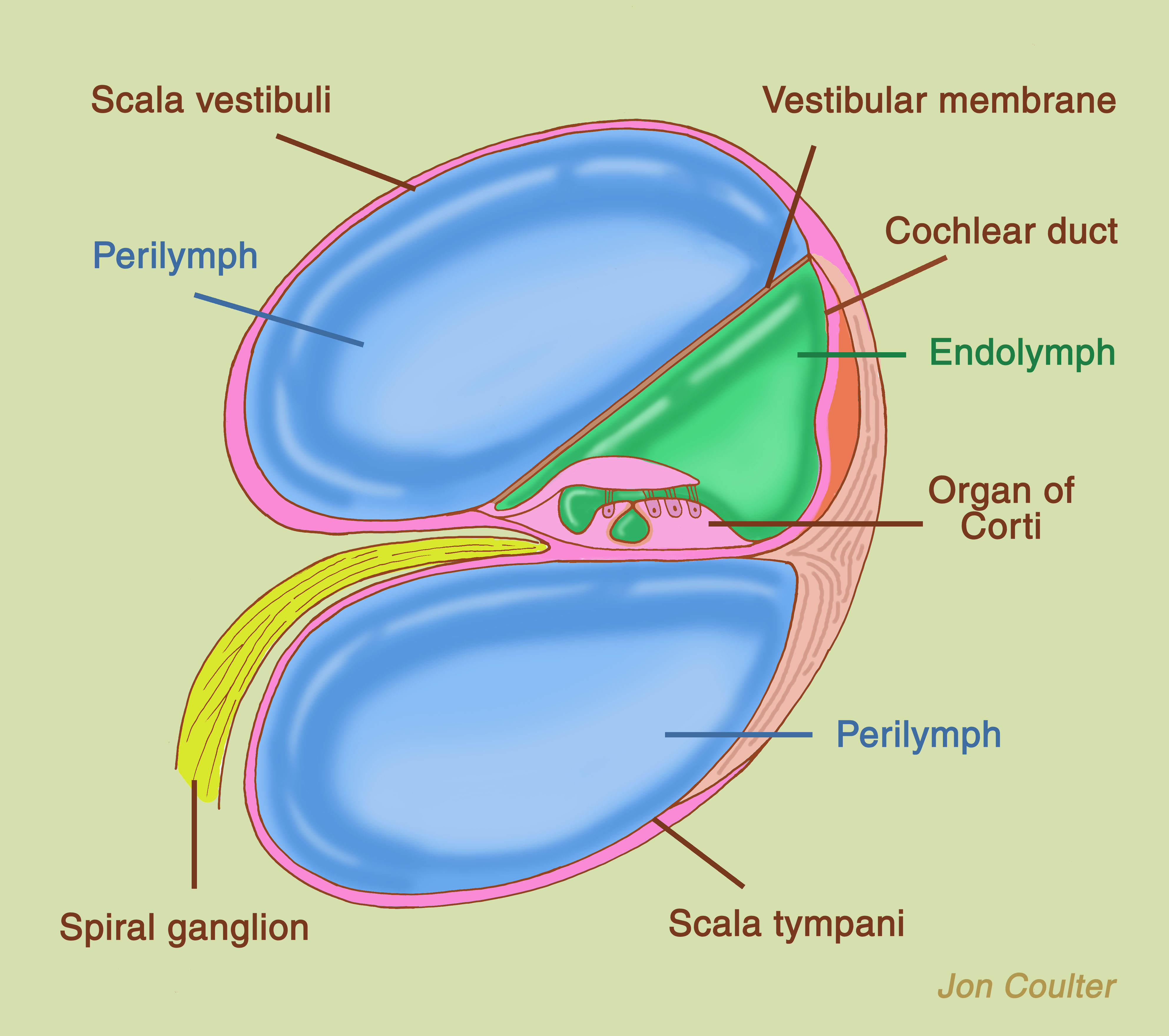
This shows a cross-section of the cochlea with the vestibular membrane, the membrane that separates endolymph and perilymph, in its proper position.
The endolymph is formed within the cochlea and circulates throughout the inner ear. An extension of the inner ear known as the endolymphatic duct and sac lays against the dura, the covering of the brain. Within the endolymphatic sac, endolymph is filtered through to the connective tissue around the sac. From here, the fluid can enter blood vessels or enter the spinal fluid space. The volume of endolymph in the inner ear is extremely small and is measured in nanoliters.
About Dr. Prasad

Dr. Sanjay Prasad MD FACS is a board certified physician and surgeon with over twenty years of sub-specialty experience in Otology, Neurotology, advanced head and neck oncologic surgery, and cranial base surgery. He is chief surgeon and founder of the private practice, Metropolitan NeuroEar Group, located in the metropolitan Washington D.C. area.
Find us on Facebook
Follow us on Twitter
- MenieresDx tweeted Our website has launched! http://t.co/71s6kHbqOctober 27, 2011 at 11:24am
- MenieresDx tweeted Visit our brand new educational website, http://t.co/JAerZc56, for more information on Meniere's disease!October 27, 2011 at 10:56am
- MenieresDx tweeted Have you been diagnosed with Meniere's disease? Need information on what it is? Want access to others with the same condition? Follow us!January 07, 2011 at 12:41pm