

Paraganglioma
A paraganglioma is a tumor that originates from cells called paraganglia found in the middle ear:*Glomus tympanicum tumors are small sized tumors originating in the middle ear.
*Glomus jugulare arise from paraganglia in or around the jugular bulb, and as they grow they occlude this venous structure.
Overview of Paraganglioma
A paraganglioma is a tumor that can originate from cells called paraganglia found in the middle ear. Other names commonly used for paraganglioma include chemodectoma, glomus tympanicum, and glomus jugulare tumors.
Tumors are classified according to whether they are benign or malignant. Benign tumors, in general, grow slowly and do not spread throughout the body, whereas malignant tumors have a faster growth pattern and can spread to other organs in the body. In most instances, paragangliomas behave as benign tumors. In very rare circumstances, these tumors can behave like malignant lesions and spread to lymph nodes in the neck.
ANATOMY
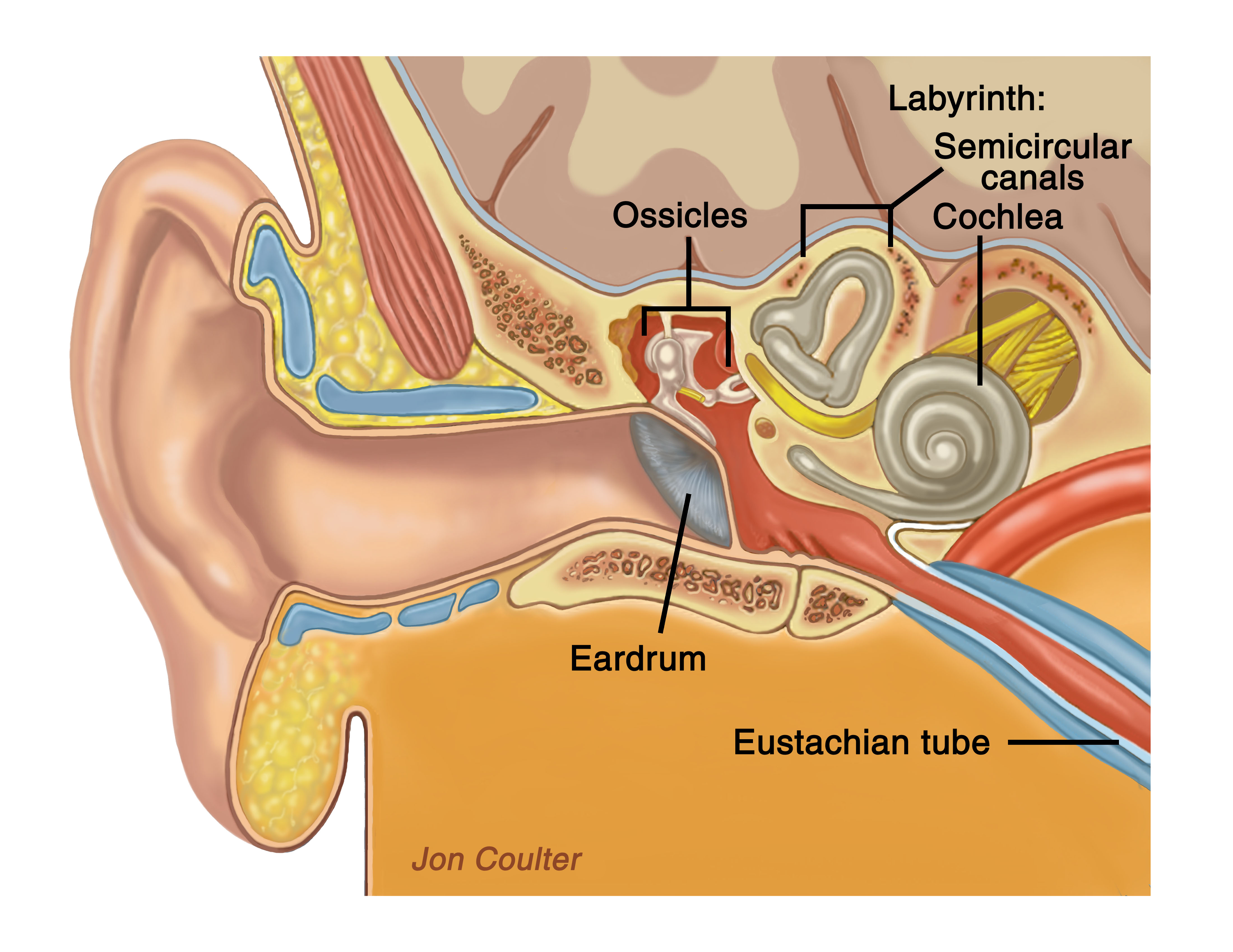
The temporal bone anatomy is shown, including the ear canal, ear drum, middle ear, and inner ear.
A brief anatomical review will allow a better understanding of the surrounding structures that can be involved with these tumors. Sound entering the ear canal causes vibrations of the tympanic membrane (eardrum). The tympanic eardrum is attached to one of three ossicles (bones) found in the middle ear called the malleus. Vibrations of the malleus bone are transmitted to another ossicle, the incus, which in turn transmits the vibrations to the smallest of the three ossicles, the stapes. Vibrations of the stapes bone are transmitted to the inner ear. The inner ear is made of a bony labyrinth filled with fluid and membranes. As the stapes vibrates, the fluid in the inner ear is set into motion. The cilia on the ends of hair cells within the inner ear are bent and an electrical signal is generated, traveling along the cochlear (hearing) nerve and then the brain. The inner ear is composed of the cochlea, which is responsible for hearing, and the semicircular canals that detect angular acceleration of the head. This acceleration data, coupled with visual data and proprioceptive (position in space) data, determine our sense of balance.
The internal auditory canal is a bony canal from the inner ear to the posterior fossa where the cerebellum and brainstem are found. It contains three different types of nerves: the hearing, balance, and facial nerve. The facial nerve is responsible for transmitting electrical impulses to the facial muscles, allowing motion of the face. This is the nerve that is responsible for our ability to raise our eyebrows, close our eyes, flare our nostrils and raise the corner of the mouth on that side. It is also responsible for conducting information regarding taste from the front 2/3 of the tongue and also producing tears from the lacrimal glands in the eyes.
The mastoid bone is a bony prominence found behind the external ear. The mastoid is composed of multiple air cells that communicate with the middle ear. Deep within the mastoid is a large vein called the sigmoid sinus that collects venous blood from that side of the brain. The sigmoid sinus empties blood into a small cavity called the jugular bulb. From there, the blood enters a large vein in the neck called the internal jugular vein. The internal jugular vein then empties into larger blood vessels leading to the heart. The jugular bulb is located in the lower part of the middle ear. Intimately associated with the jugular bulb are several nerves that are involved with the swallowing mechanism. These nerves are also responsible for the proper functioning of the vocal cord. Paragangliomas can thus alter the quality of voice and the swallowing mechanism.
Glomus Tympanicum
Paraganglioma or glomus tumors are named according to where they originate. Glomus tympanicum tumors are small sized tumors originating in the middle ear. They generally do not involve the jugular bulb but can at times erode the bone over the jugular bulb. The symptoms that lead to this diagnosis include pulsatile tinnitus (ringing in the ears) that is heard with each heartbeat.

Here, a glomus tympanicum is seen pulsating in the middle ear.
Glomus Jugulare tumors
These tumors arise from paraganglia in or around the jugular bulb. As they grow, they occlude this venous structure. With additional growth, venous blood from the brain also gradually begins to shift toward the other sigmoid sinus and jugular bulb. Symptoms of these tumors include pulsatile tinnitus (ringing in the ears) that is heard with each heartbeat.
Here, a glomus jugulare is seen pulsating in the jugular bulb.
Carotid Body Tumors
Carotid body tumors arise from paraganglia located within the covering of the carotid artery in the neck. The initial symptoms can be pain and a pulsating mass in the neck.
About Dr. Prasad

Dr. Sanjay Prasad MD FACS is a board certified physician and surgeon with over twenty years of sub-specialty experience in Otology, Neurotology, advanced head and neck oncologic surgery, and cranial base surgery. He is chief surgeon and founder of the private practice, Metropolitan NeuroEar Group, located in the metropolitan Washington D.C. area.